
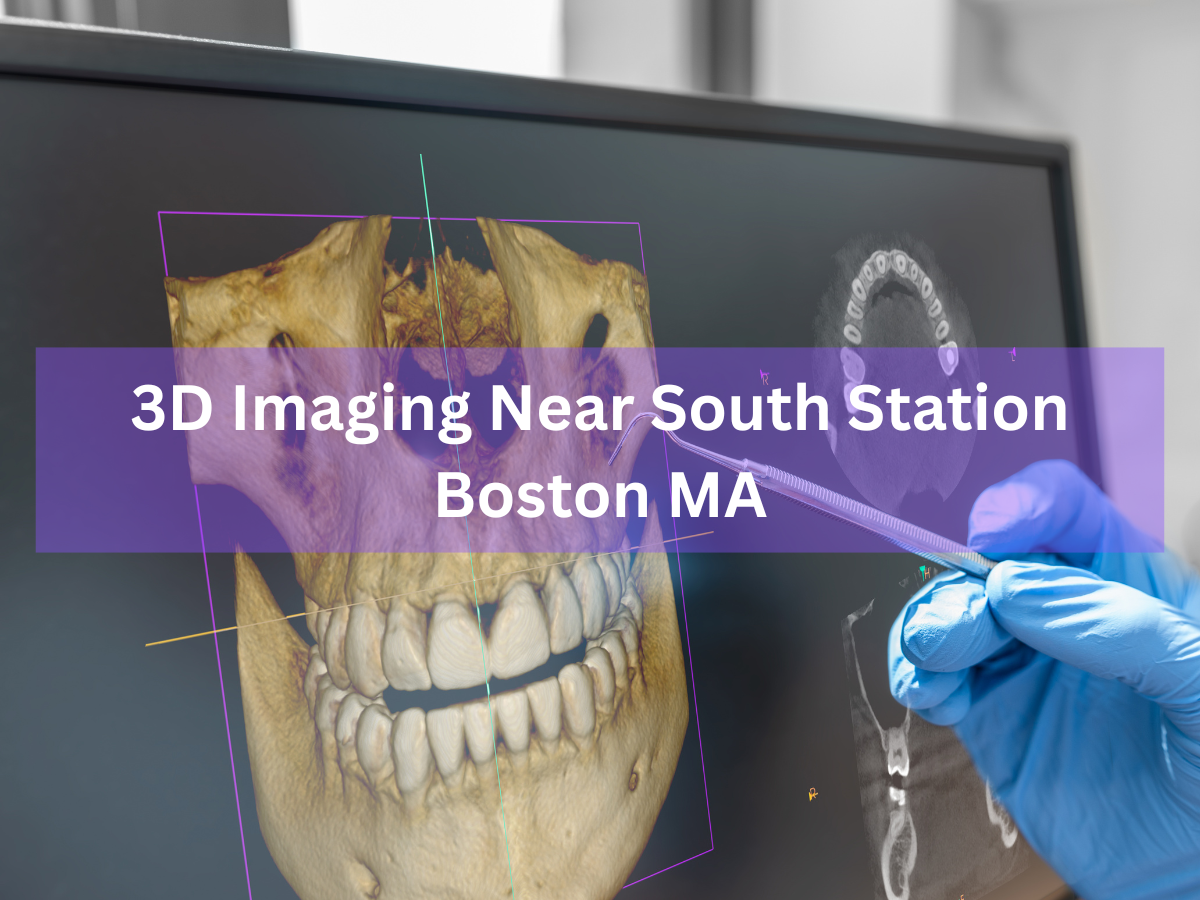
A state-of-the-art 3D dental scan being performed at Congress Dental Group in Boston. Located near Boston’s South Station transit hub, Congress Dental Group offers cutting-edge 3D dental imaging technology to improve diagnosis and treatment planning. Unlike a conventional 2D X-ray, a three-dimensional cone-beam CT (CBCT) scan captures a complete volume of your teeth, jawbone, and surrounding tissues in a single session. This means our team can see detailed views of tooth roots, nerve canals, sinus cavities and more – information that flat 2D X-rays often miss. By using CBCT in our South Station office, dentists at Congress Dental can plan procedures (such as implants or wisdom tooth extractions) with unprecedented accuracy. The result for patients is often fewer office visits, improved safety, and more confident treatment outcomes.
What is 3D Dental Imaging (Cone Beam CT)?
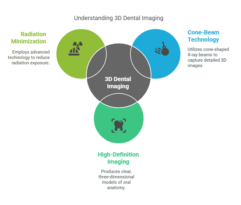
3D dental imaging refers to specialized X-ray technology called cone-beam computed tomography (CBCT) that produces a volumetric (3D) model of the patient’s oral anatomy. RadiologyInfo explains that a dental cone-beam CT is “a special type of X-ray equipment” used when regular dental X-rays are not sufficient. It can generate “three-dimensional (3-D) images of dental structures, soft tissues, nerve paths and bone in the craniofacial region in a single scan”. In practice, the patient sits or stands and a cone-shaped X-ray beam rotates around the head, capturing multiple images from every angle. A computer then reconstructs these into a high-definition 3D image. Importantly, modern dental CBCT units like the CS 9300 used at Congress Dental are designed to minimize radiation. For example, Boston University notes their i-CAT CBCT scanner delivers high-definition 3D images “with substantially less radiation to the patient compared to a regular CT examination”. In short, 3D dental imaging gives your dentist a comprehensive internal view of your mouth and jaw in three dimensions, helping ensure nothing is overlooked during diagnosis or planning.
How Does 3D Dental Imaging Work?
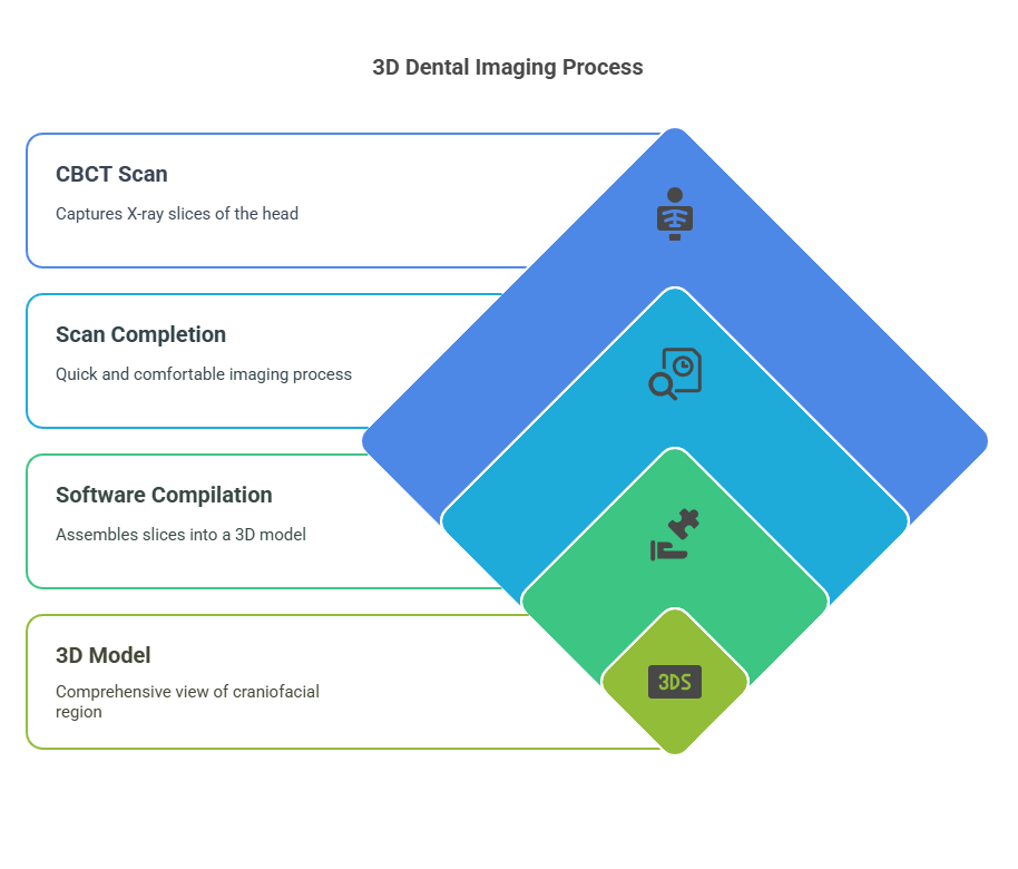
Performing a 3D dental scan is fast and painless. You will be seated or standing still while the CBCT machine gently rotates around your head to collect images. As Ace Dental Boston describes, “the ConeBeam CT imaging system is a digital X-ray scanner that rotates once around your head” in about 20 seconds. During this quick sweep, the machine takes many X-ray “slices” of the entire craniofacial region. It is essentially like taking hundreds of X-rays in one pass. Modern CBCT devices often complete a full scan in only 10–20 seconds. Once the scan is finished, the imaging software compiles all the slices into a 3D model. Dentists can then virtually “slice” this model in any direction (axial, coronal, or sagittal) and even view it in color. As one Boston practice notes, this technology allows “virtually unlimited views of your teeth, face, and neck with just one scan,” in full 3D. The entire process is quick, easy and extremely comfortable – there’s no pain or confined spaces involved.
Benefits of 3D Imaging vs Traditional X-Rays
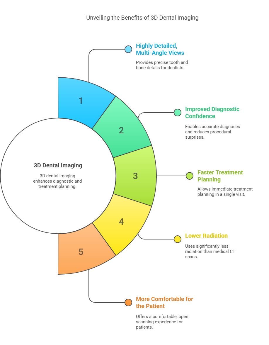
3D dental imaging offers significant advantages over conventional 2D X-rays. For example, Congress Dental Group highlights that their CBCT system provides “precise, crystal-clear digital images while minimizing exposure to radiation”. Key benefits include:
-
Highly detailed, multi-angle views: 3D scans reveal the exact shape and orientation of each tooth and bone. Dentists can inspect any hidden issues (like tiny fractures or early infection) that a flat X-ray might miss. High-resolution CBCT data lets clinicians “see your teeth with unprecedented detail”.
-
Improved diagnostic confidence: With a full 3D map of your mouth, dentists can make more accurate diagnoses and avoid surprises during procedures. For example, they can pinpoint the exact location of nerves and sinuses, reducing the risk of complications. Congress Dental notes that their 3D system allows them to perform a wider range of diagnoses and even share images with other doctors to coordinate care.
-
Faster treatment planning: Because the scan is done in-office and the 3D model is available immediately, you may leave the appointment with a complete treatment plan. There’s no waiting for off-site imaging. In fact, one practice says CBCT enables “immediate virtual diagnosis and treatment planning in one visit”.
-
Lower radiation (focused on area of interest): Although a 3D scan uses more radiation than a single bitewing X-ray, it is focused on a specific area and uses far less radiation than a full medical CT scan. Congress Dental follows the ALARA (As Low As Reasonably Achievable) principle by targeting only the necessary field of view, further reducing exposure. For example, CBCT machines typically deliver 10–30 times less radiation than a hospital CT for the same region. The result is a safer exam for the patient while obtaining CT-like detail.
-
More comfortable for the patient: Most dental CBCT units are open and allow you to stand or sit during the scan. There are no claustrophobic tunnels or sedation required. The system’s design accommodates patients of all sizes comfortably.
In summary, 3D CBCT scans pack more clinical information into a single, quick scan. Congress Dental Group invested in this technology precisely to enhance patient care – providing sharper images (high resolution), faster results (immediate planning), and a comfortable scanning experience, all with minimal radiation.
Common Uses of 3D Dental Imaging
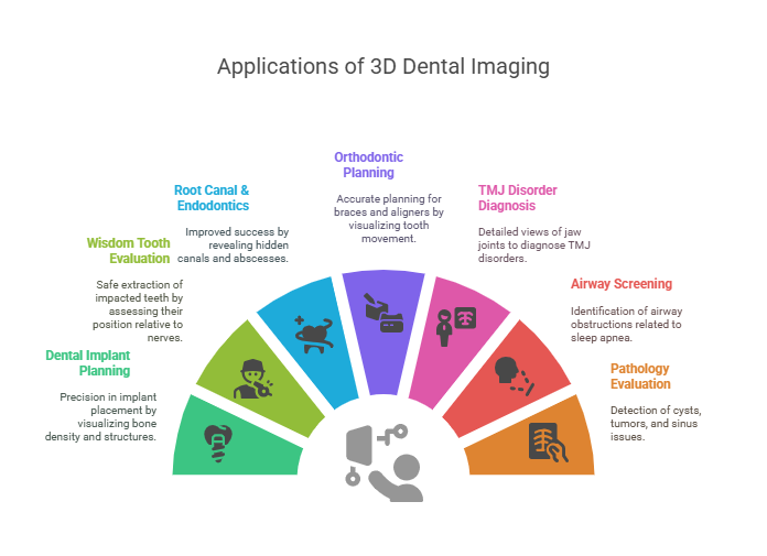
Dentists and specialists use 3D dental imaging for many advanced procedures. Some of the most common applications include:
-
Dental Implant Planning: CBCT gives a clear view of bone density and vital structures so implants can be placed with millimeter precision. Surgeons can avoid nerves and sinuses and determine the optimal implant size and angle.
-
Wisdom Tooth & Impacted Tooth Evaluation: The exact position of an impacted tooth relative to adjacent teeth and nerves is easily seen in 3D, aiding safe extraction. Oral surgeons rely on CBCT for complicated extractions.
-
Root Canal & Endodontics: 3D scans reveal hidden canals, cracks, or abscesses that might not show on 2D X-rays, improving endodontic success.
-
Orthodontic Diagnosis & Planning: Orthodontists use CBCT to assess jaw relationships and tooth alignment in three dimensions. It helps in planning braces or clear aligners by showing how teeth will move through bone.
-
TMJ and Jaw Joint Disorders: 3D imaging provides coronal, sagittal and axial views of the temporomandibular joint. This helps diagnose TMJ disorders by showing joint anatomy and bone position in detail.
-
Airway and Sleep Apnea Screening: CBCT can visualize airway space and nasal passages, which is useful in identifying obstructions related to sleep apnea or breathing problems.
-
Sinus and Pathology Evaluation: Any cysts, tumors, or sinus issues are visible in a 3D CBCT scan, along with bone lesions or fractures. For instance, CBCT is used to measure jaw tumors and their impact on surrounding structures.
In short, whenever a detailed view of the teeth, jaws or facial bones is needed, 3D dental imaging is invaluable. The American Academy of Oral and Maxillofacial Radiology notes that cone beam CT is used for implant placement, impacted teeth, TMJ, assessing the jaw and sinuses, and more. By contrast, regular X-rays or panoramic films give only a flat picture, which can miss critical information.
3D Imaging at Congress Dental Group (Boston, South Station)
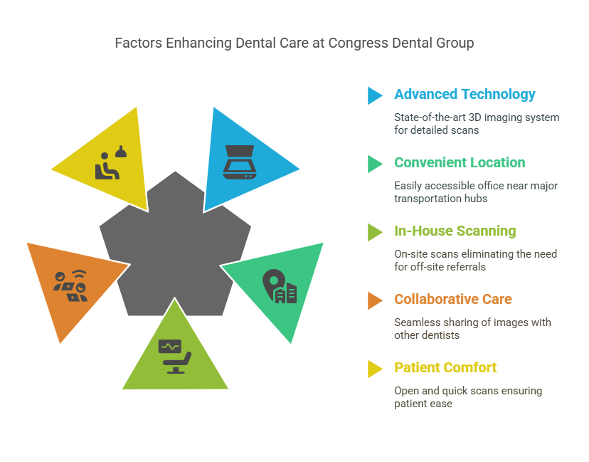
Congress Dental Group brings this advanced technology right into the heart of downtown Boston. Our Financial District office on 160 Federal Street, just steps from South Station, is equipped with a state-of-the-art CS 9300 3D imaging system (Cone Beam CT). Dr. Robert Page and our team use this device for on-site 3D scans, meaning patients do not need to be referred off-site or to a hospital. With our in-house scanner, you can have the CBCT exam and discuss the results with your dentist in one visit.
Our CS 9300 CBCT unit provides exceptionally detailed images with sub-millimeter accuracy. As our website notes, this system delivers unmatched visualization of anatomical detail, helping us explain treatment plans and share data easily. In fact, Congress Dental’s 3D software allows us to “quickly and easily share 3D images of the area of concern with your general dentist,” which helps collaborating doctors “improve your experience and deliver a positive treatment outcome”. In other words, we use 3D imaging not only for better diagnostics, but also to enhance coordination with any specialists you may need.
Because our office is so accessible (near the Broadway and Red Line trains), patients from all over Boston can benefit from this technology without traveling far. If you work or live near South Station, Faneuil Hall, or the Financial District, you can easily schedule your CBCT scan at Congress Dental Group. We strive to provide every patient with the best technology and comfort – which includes open, quick 3D scans.
What to Expect During Your 3D Dental Scan
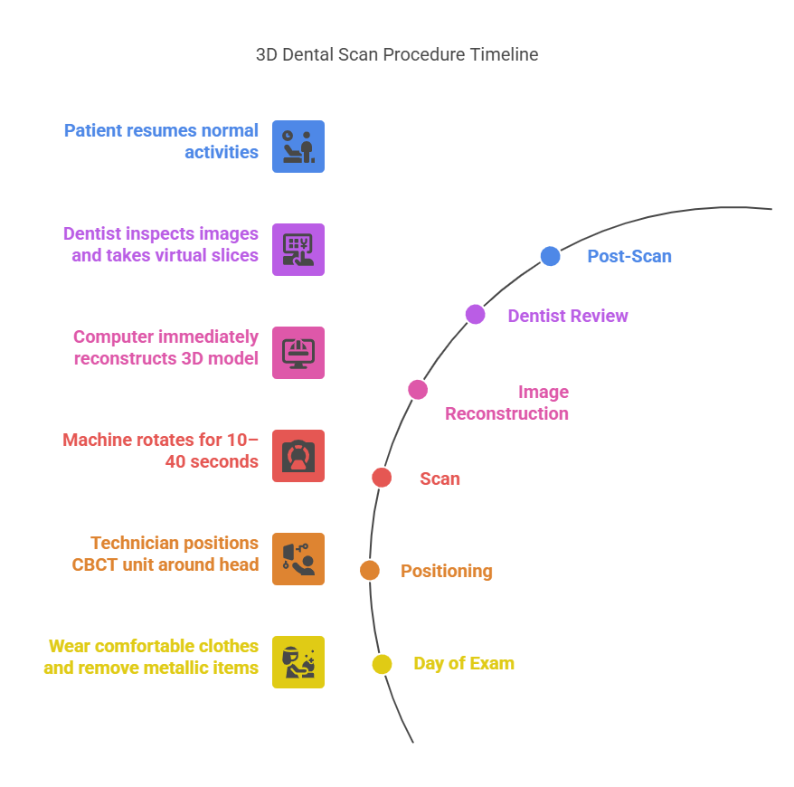
Getting a 3D dental scan at our office is simple and quick. There is little to no special preparation needed. On the day of your exam, just wear comfortable clothes and remove any metallic items (like jewelry or glasses) that could block the X-rays. You will either sit or stand next to the scanner and our technician will position the CBCT unit around your head. Then you’ll be asked to stay very still, and the machine will rotate for about 10–40 seconds (depending on the scan settings). You may hear a whirring noise, but you won’t feel anything – it is a painless, non-invasive procedure.
Because the scan is so fast, most patients are done in less than a minute. Once the images are captured, the computer reconstructs the 3D model immediately. The dentist can then inspect the images on screen, zooming in on any area and taking virtual “slices” as needed. After the scan, you can eat and resume normal activities right away. The whole process is safe and comfortable.
Safety and Radiation in Dental 3D Imaging
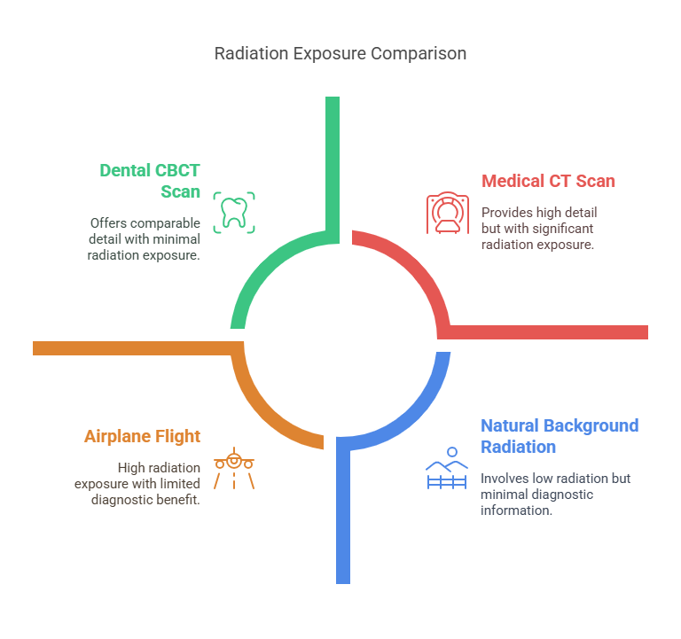
Safety is a top priority in any imaging procedure. Dental cone-beam CT does involve X-ray exposure, but when used properly it is very low. Because CBCT units like our CS 9300 focus on a limited area (just the mouth and jaw), the radiation dose is significantly lower than a traditional medical CT scan, yet provides comparable detail. In fact, Boston University reports that their dental CBCT has “substantially less radiation to the patient compared to a regular CT examination”. Moreover, modern scanners follow ALARA (As Low As Reasonably Achievable) principles. Congress Dental’s system lets us choose a small field of view to expose only the area of concern, drastically reducing unnecessary exposure.
For context, a dental CBCT scan typically exposes you to about the same amount of radiation as a few days of natural background radiation, or a couple weeks of flying on an airplane – far less than a medical head CT. We always take precautions such as using lead aprons/neck collars and limiting scans to necessary cases. If you are pregnant, we will discuss the risks (as with any X-ray) and may postpone elective imaging. Rest assured that when recommended by your dentist, the benefit of accurate diagnosis far outweighs the minimal risk. Every scan at our Boston office is performed by trained professionals, ensuring the safest possible imaging.
Cost and Insurance Coverage
3D imaging is a powerful diagnostic tool, but patients often wonder about cost. Insurance coverage for cone-beam CT scans varies. In many cases, dental plans or medical insurance will cover 3D scans when they are deemed medically necessary (such as for implant planning or to investigate pathology). Some plans consider CBCT an “alternate radiographic” exam. At Congress Dental Group, we bill most insurances for eligible procedures and can help you check your benefits ahead of time. Often the expense can be bundled with treatment fees (for example, implant placement). If insurance does not cover it, we offer financing options. Ultimately, we believe the precision gained from 3D imaging – leading to better outcomes and fewer complications – is well worth the investment in your dental health.
People Also Ask
Q: What is 3D dental imaging?
A: 3D dental imaging (cone-beam CT) is an advanced X-ray technique that produces a three-dimensional view of the teeth, jaw, and face in a single scan. It provides more detailed information than regular flat X-rays by showing depth and structure, which helps dentists diagnose problems and plan treatments more accurately.
Q: How long does a 3D dental scan take?
A: A modern 3D CBCT scan is very fast. The machine usually rotates around your head in about 10–20 seconds and captures all the data in one go. Including setup, the entire visit might take only a few minutes.
Q: Is 3D dental imaging painful?
A: No, a 3D dental scan is completely painless. You simply sit or stand still while the machine rotates around you. There are no needles, no discomfort, and the procedure is non-invasive.
Q: How much radiation does a 3D dental X-ray use?
A: Dental CBCT uses more radiation than a single traditional X-ray, but much less than a medical CT scan. For example, our CBCT unit delivers crystal-clear images while “minimizing your exposure to radiation”, and BU notes this is “substantially less” than a regular CT. Overall, the dose is quite low and focused only on the area of interest.
Q: Why do dentists use 3D imaging?
A: Dentists use 3D imaging because it reveals hidden details that 2D X-rays cannot show. For instance, it can reveal the exact location of nerves, the density of jawbone for implants, or problems in sinuses. This leads to safer and more effective treatment plans. Congress Dental Group uses 3D imaging to increase diagnostic confidence and help patients avoid surprises during procedures.
Q: Where can I get a 3D dental scan near South Station, Boston?
A: You can get a 3D dental scan at Congress Dental Group’s Federal Street office, which is right next to South Station. We have the in-house CBCT technology and can perform the scan during your dental visit.
Q: How does 3D imaging benefit dental implants?
A: Before placing an implant, dentists need to know exactly how much bone is available and where key structures (like nerves) lie. A 3D scan provides a virtual model for precise implant planning. This minimizes risks (e.g. nerve injury) and often speeds up healing by getting it right the first time.
Q: Are 3D dental scans covered by insurance?
A: Coverage varies by plan. In many cases, if the scan is needed for a covered procedure (like implant or oral surgery), insurance will pay for it. Congress Dental staff can assist in verifying coverage. Even if not fully covered, financing options are usually available.
Q: Can children have 3D dental imaging?
A: Yes, but dentists use it only when necessary. Because children are more sensitive to radiation, CBCT scans in pediatric patients are done only if the detailed 3D view is crucial to treatment. Otherwise, standard X-rays (with even lower radiation) are used.
Q: How does a cone beam CT differ from a regular CT?
A: Cone-beam CT (dental CBCT) is specifically designed for teeth and jaws. It uses a cone-shaped beam and takes just one quick rotation to gather images. It produces high-quality 3D images of dental structures with far less radiation and a lower cost than a medical CT scanner. RadiologyInfo notes that cone beam CT produces similar types of images as conventional CT but in a smaller, more compact machine.
Frequently Asked Questions (FAQs)
Where is Congress Dental Group’s South Station office located?
Congress Dental Group’s Boston downtown office is at 160 Federal Street, 1st Floor, Boston, MA 02110, just a block from South Station. Our central location makes us easily accessible by train, bus, or car, and we serve the Financial District and Seaport communities.
What 3D imaging technology does Congress Dental use?
We use the Carestream CS 9300 cone-beam CT scanner – a state-of-the-art 3D imaging system. This unit provides high-resolution scans of your mouth and jaw, and can even capture a 3D cephalometric (full skull) view. The CS 9300’s flexibility and clarity help our dentists plan treatments with confidence.
Why is 3D imaging important for dental implants?
3D imaging allows us to map out exactly where to place implants. It shows the quality and quantity of bone, and reveals the precise location of nerves and sinuses. With this information, we can virtually plan the implant position before surgery, greatly improving safety and success rates.
How do I prepare for a 3D dental scan?
Preparation is minimal. You may be asked to remove jewelry, glasses, or removable dental appliances. Wear something comfortable (no metal collars). Otherwise, just come in and we’ll guide you through the brief scanning process.
How does 3D imaging improve orthodontic treatment?
For braces or Invisalign, 3D scans show the exact positions of teeth and jaws in all dimensions. This helps orthodontists diagnose bite issues more precisely and plan movement of teeth more effectively.
Do I need to remove braces or metal braces for a 3D scan?
Traditional metal braces can cause some artifacts in CBCT images, but often the images are still diagnostic. In many cases, orthodontic appliances do not have to be removed for a scan. It depends on what the scan is for; your dentist will advise you.
How often should I get a 3D dental scan?
3D scans are used only when needed – for example, before certain surgeries or complex procedures. They are not part of routine check-ups. We follow guidelines to avoid unnecessary exposure, so you will only get a CBCT if it provides clear benefit over regular X-rays.
Can 3D dental imaging detect cavities?
3D CBCT is excellent for showing teeth and bone structure, but it is not typically used to find cavities. Standard bitewing and bitewing X-rays remain the best tool for detecting tooth decay. CBCT is more often used for hard tissue assessment (bone, roots, sinuses) rather than soft issues like small cavities.
What is the cost of a 3D dental scan at Congress Dental?
The cost varies depending on the type of scan (e.g. small field vs full jaw). Generally, CBCT scans range from a few hundred dollars. We can provide a detailed estimate and will help you check your insurance benefits. The investment in precise diagnostics usually pays off by preventing more costly problems later.
How do I schedule a 3D imaging appointment?
Simply call Congress Dental Group at (617) 574-8700 or use our online booking system. Mention that you need 3D imaging (cone-beam CT) at our South Station office, and we’ll schedule a convenient time for your scan.
Conclusion
3D dental imaging has transformed modern dentistry by offering safer, faster, and more precise diagnostic capabilities than ever before. At Congress Dental Group near South Station in Boston, we proudly offer in-house cone-beam CT scanning to ensure that our patients receive the highest standard of care. Whether you’re preparing for a dental implant, assessing jaw pain, or simply looking for a more thorough diagnosis, our advanced imaging technology helps us provide more accurate treatment plans and better long-term outcomes.
If you’re seeking expert dental care in downtown Boston with access to the latest imaging tools, schedule your visit with Congress Dental Group today – and experience the confidence that comes with precision dentistry. Dr. Robert Page uses the CS 9300 3D Imaging device to diagnose potential issues more accurately and provide treatment with unprecedented confidence. We invite you to call us at (617) 574-8700 to learn more about 3D imaging in Boston, Massachusetts with our dentists at Congress Dental Group.